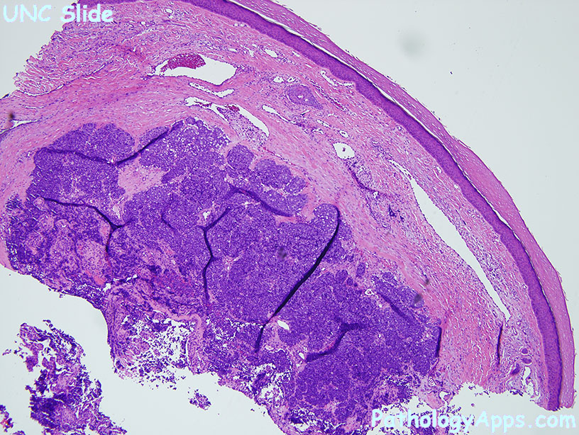
Merkel Cell Histology
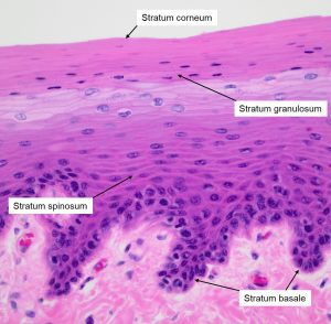
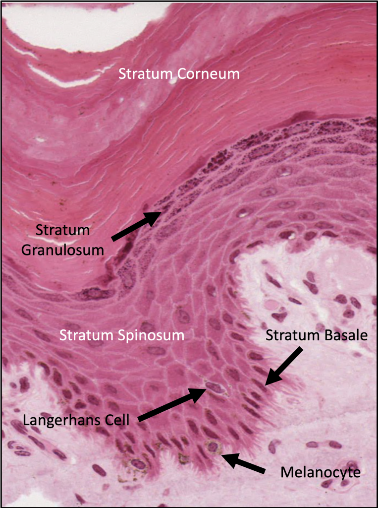
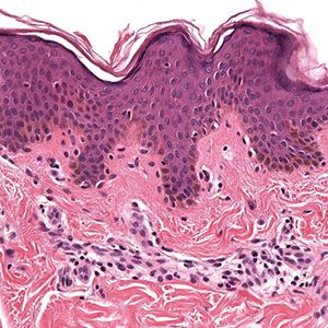
Outer layer of the skin.
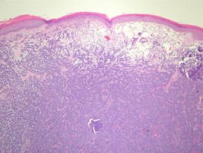
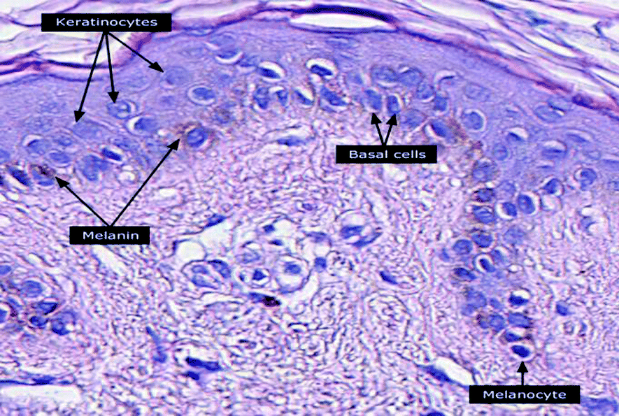
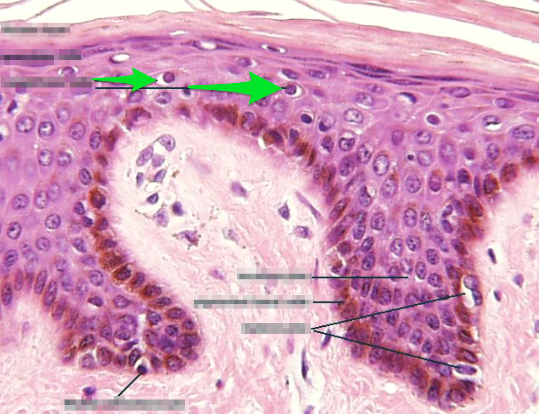
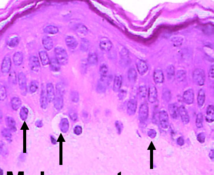
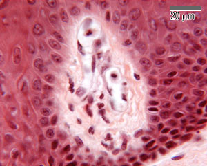
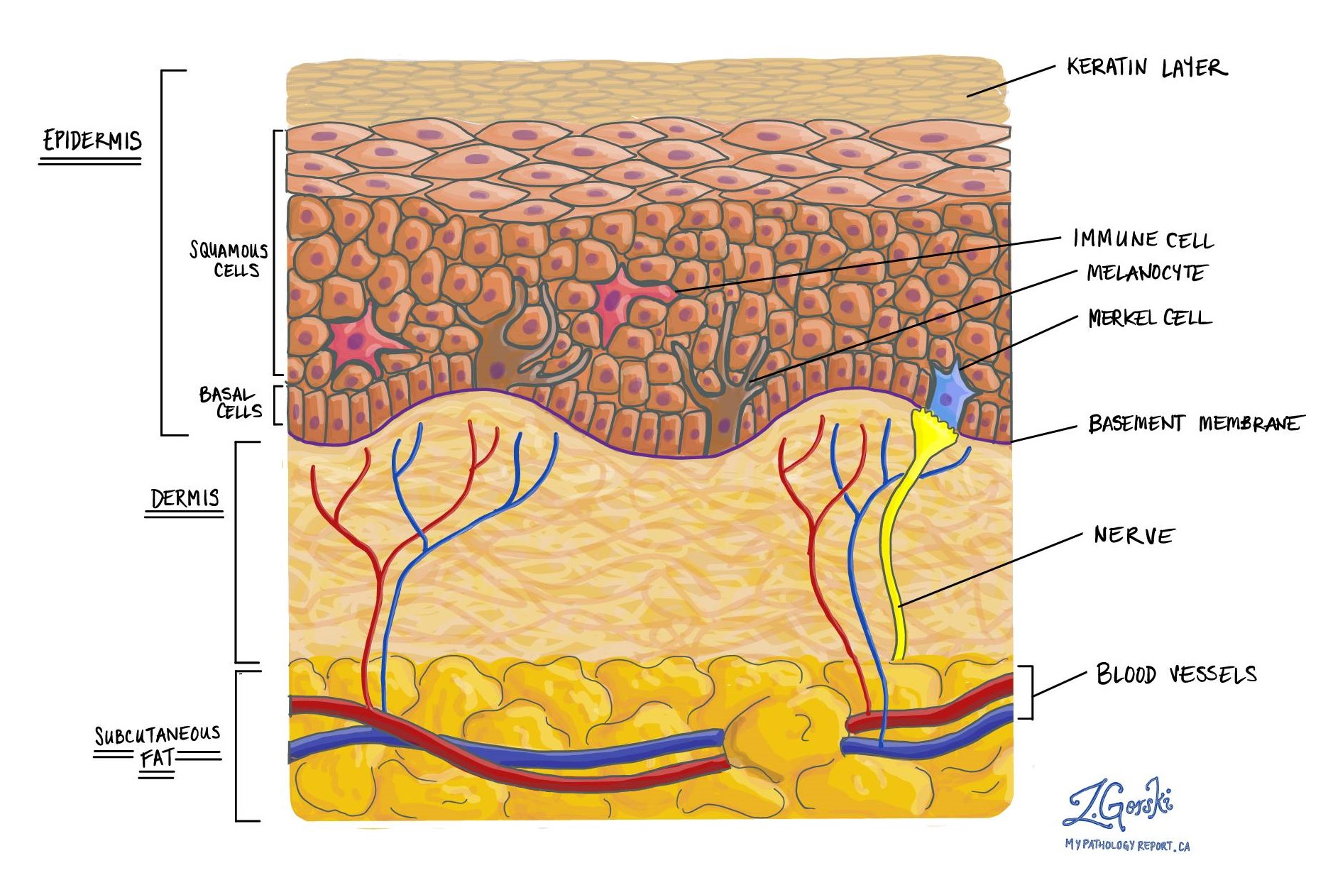
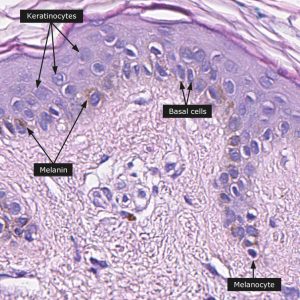
Merkel cell histology. They are difficult to tell apart from melanocytes. Merkel cells are located deep in the top layer of skin. However recent research suggests that it is unlikely that mcc originates directly from normal merkel cells. They were originally described in the late 1800s by friedrich merkel a german anatomist.
He found these cells at high density in the paws of rats and surmised they may serve as touch cells. Merkel cells are connected to nerves signaling touch sensation as touch receptors mcc was named after merkel cells due to the similar microscopic features. Merkel cells also known as merkel ranvier cells or tactile epithelial cells are oval shaped mechanoreceptors essential for light touch sensation and found in the skin of vertebrates. Merkel cells these are granular basal epidermal cells attached to a free non myelinated nerve ending which are sensitive to touch mechanoreceptors.
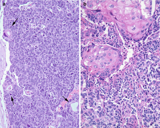
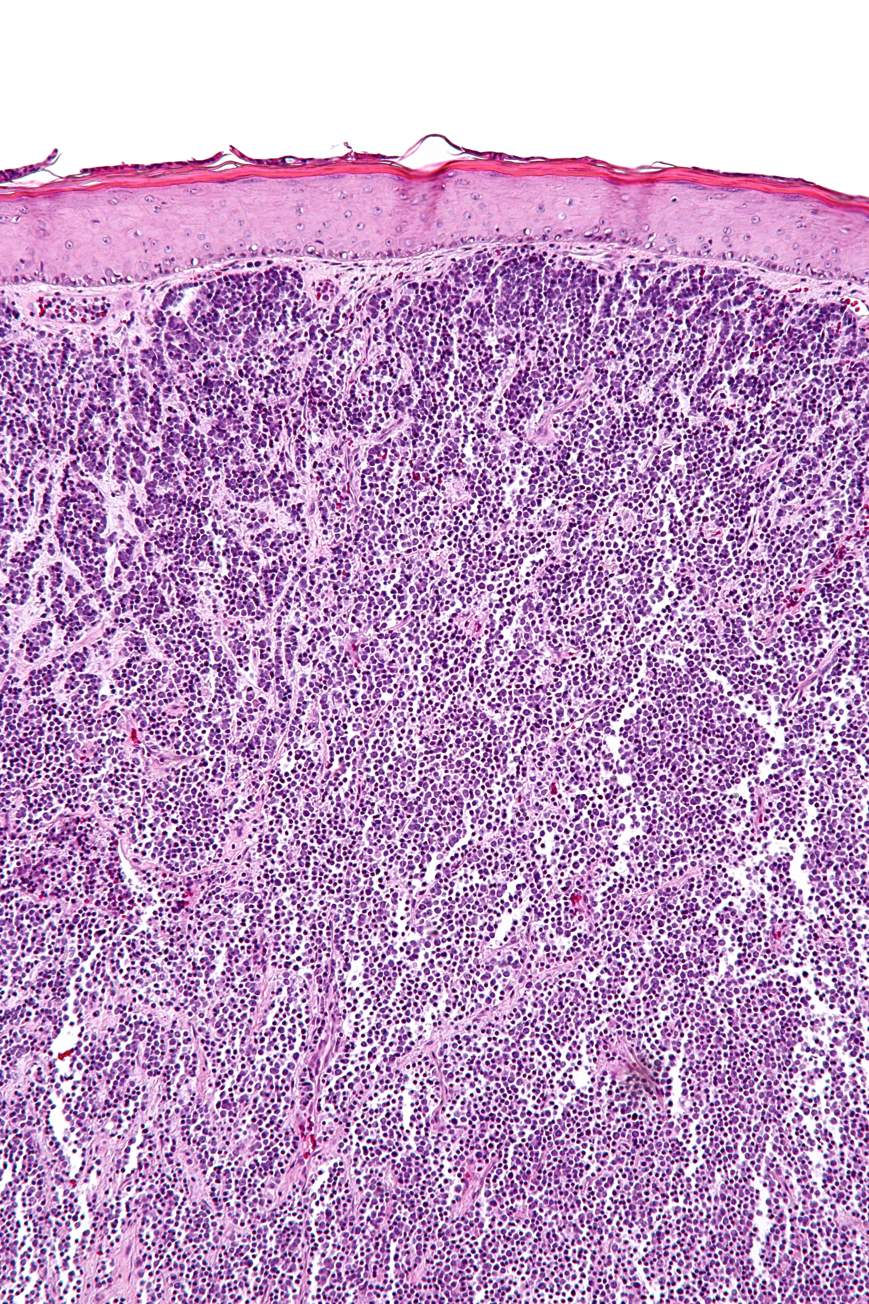
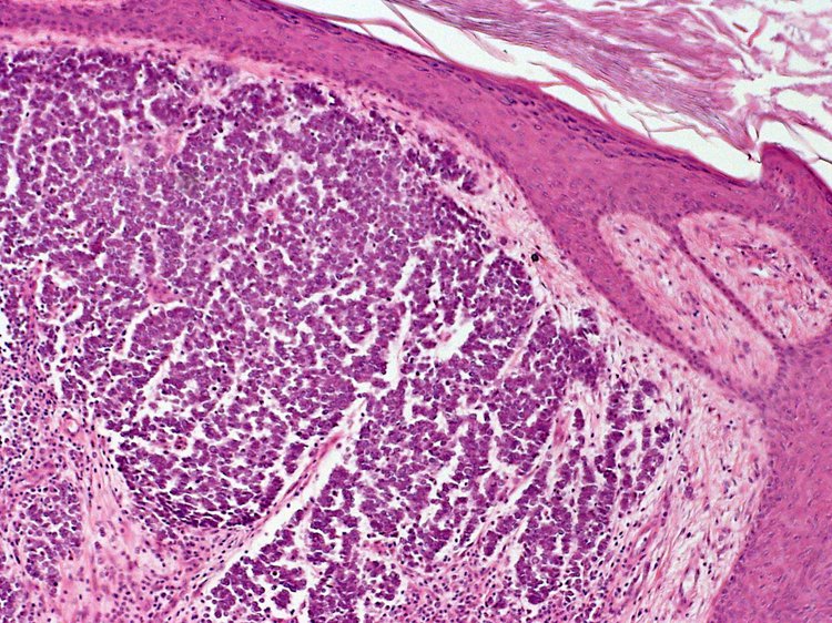
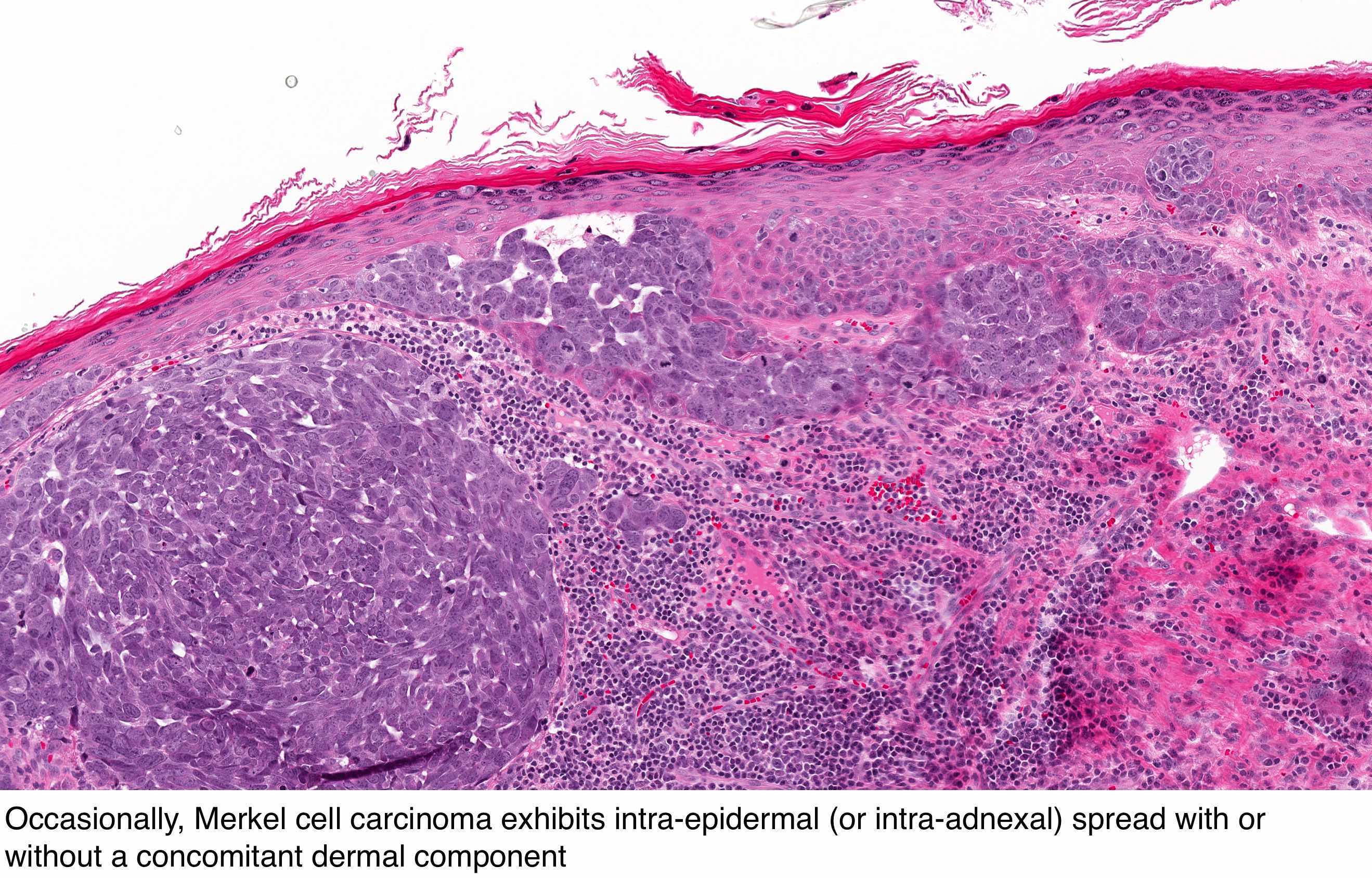
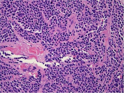
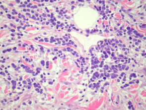
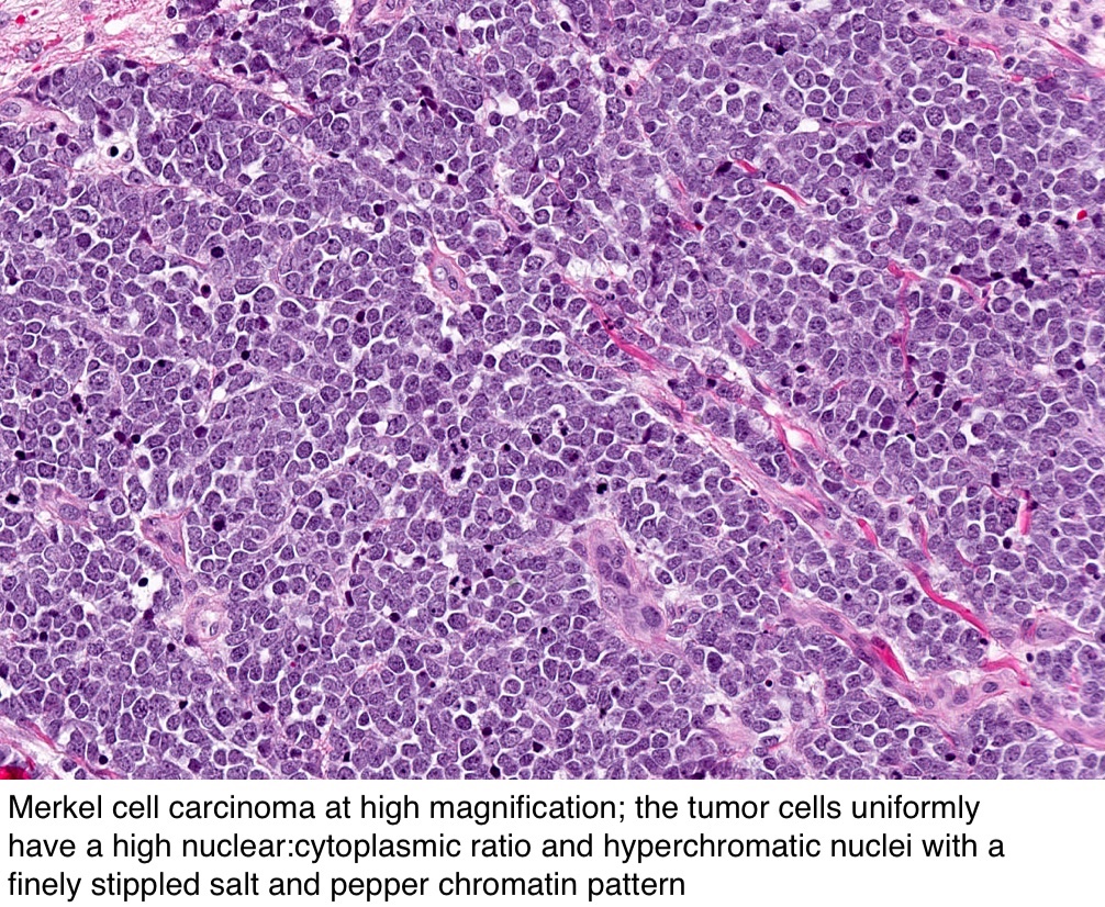
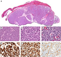
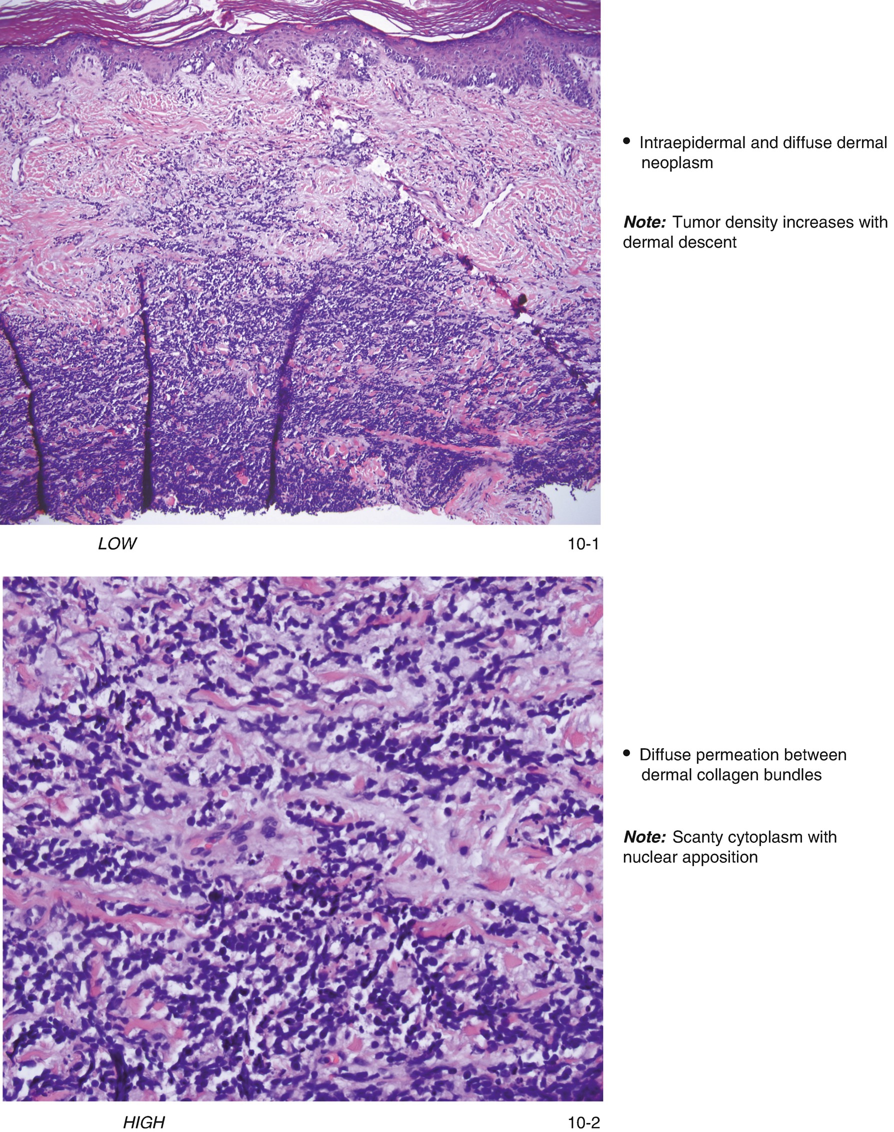
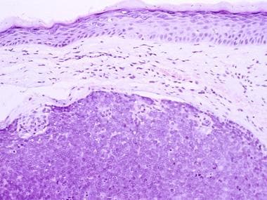
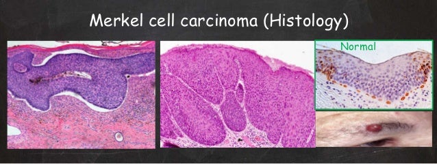
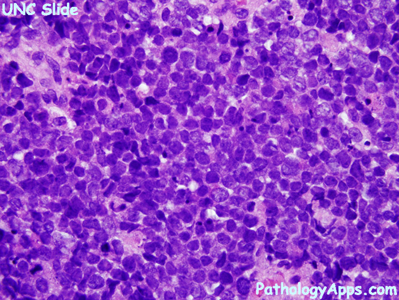
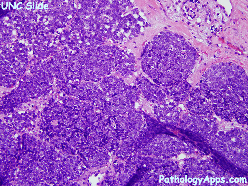
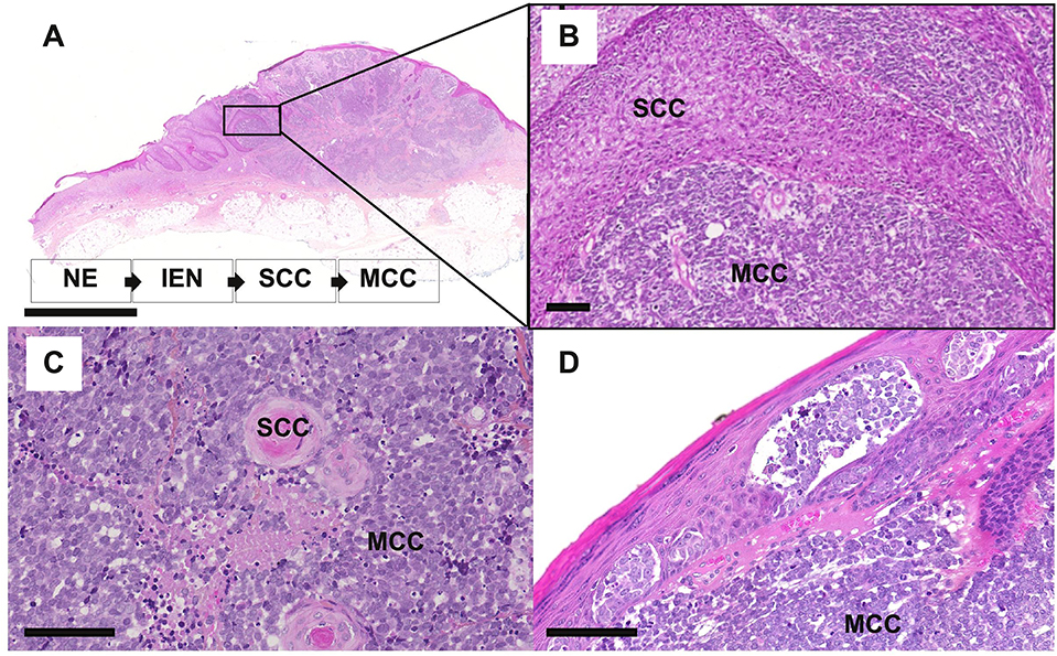
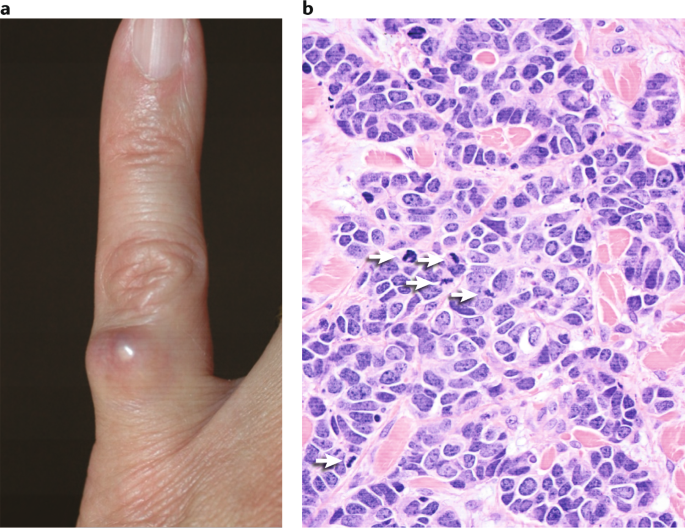
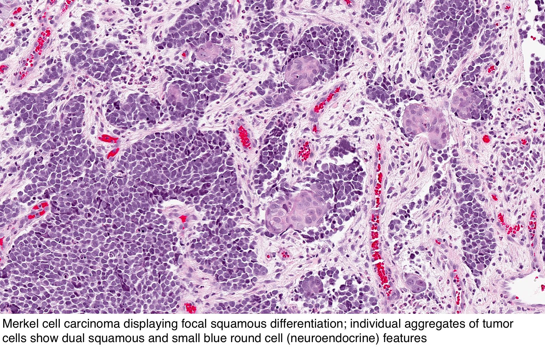
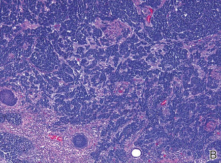
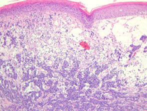
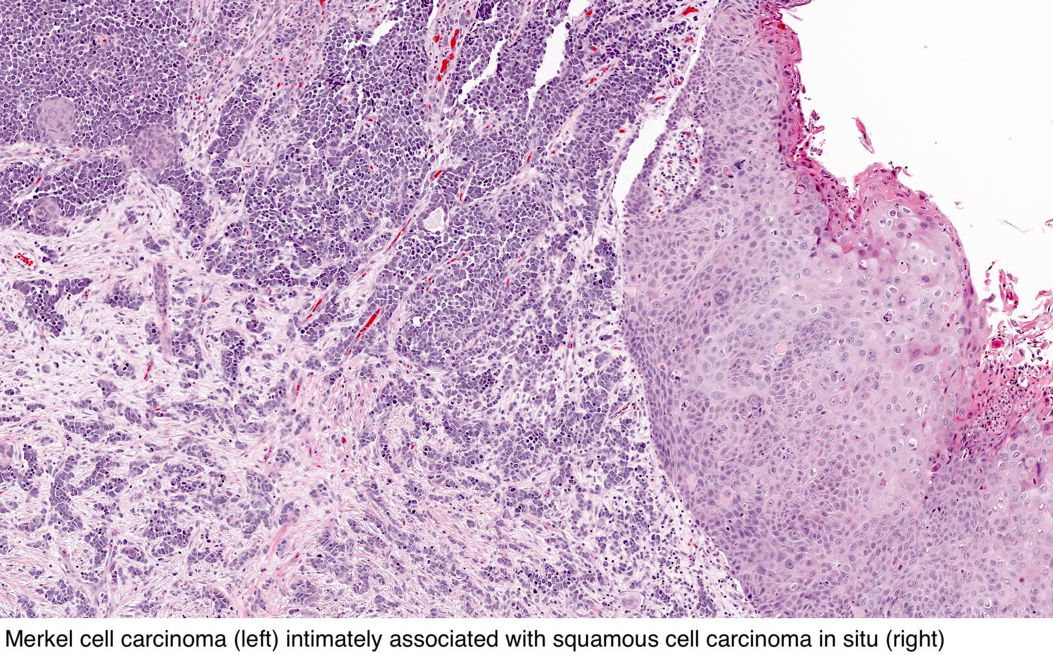
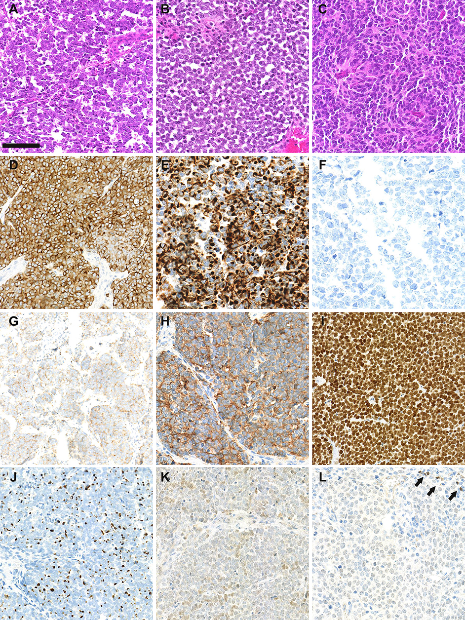
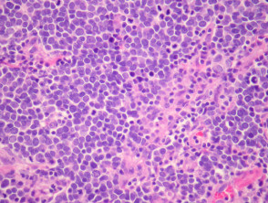
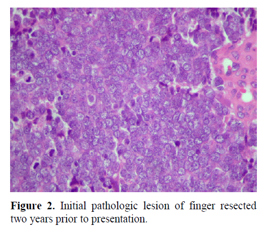
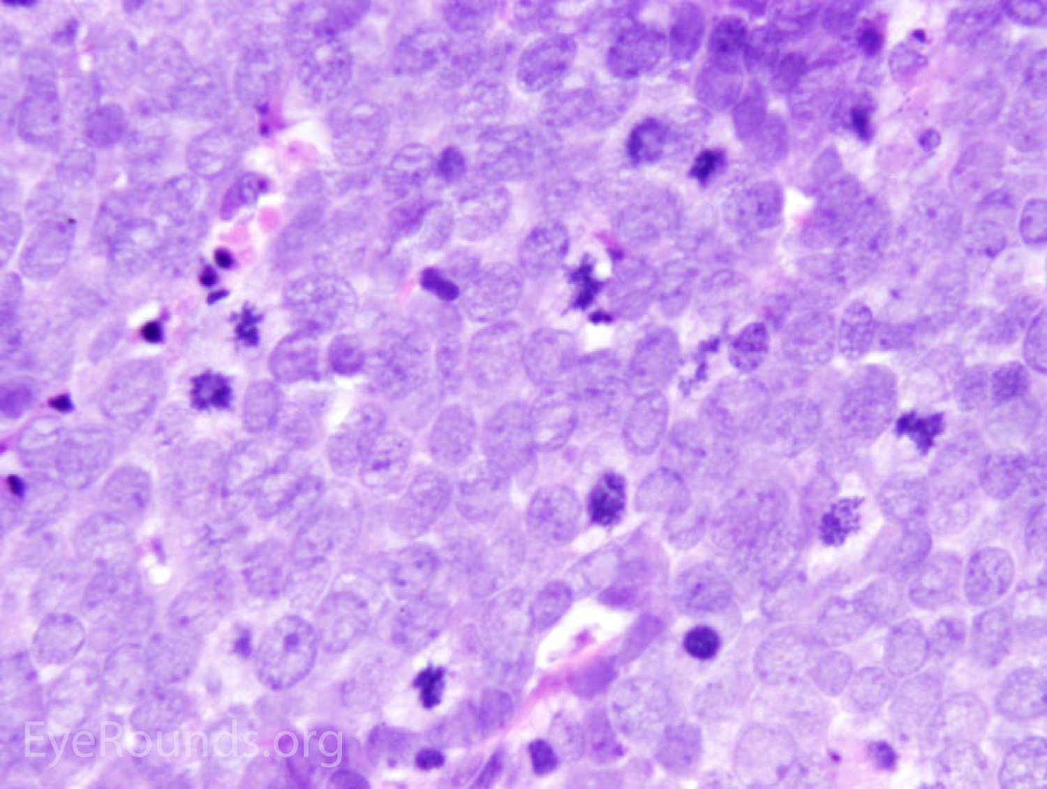
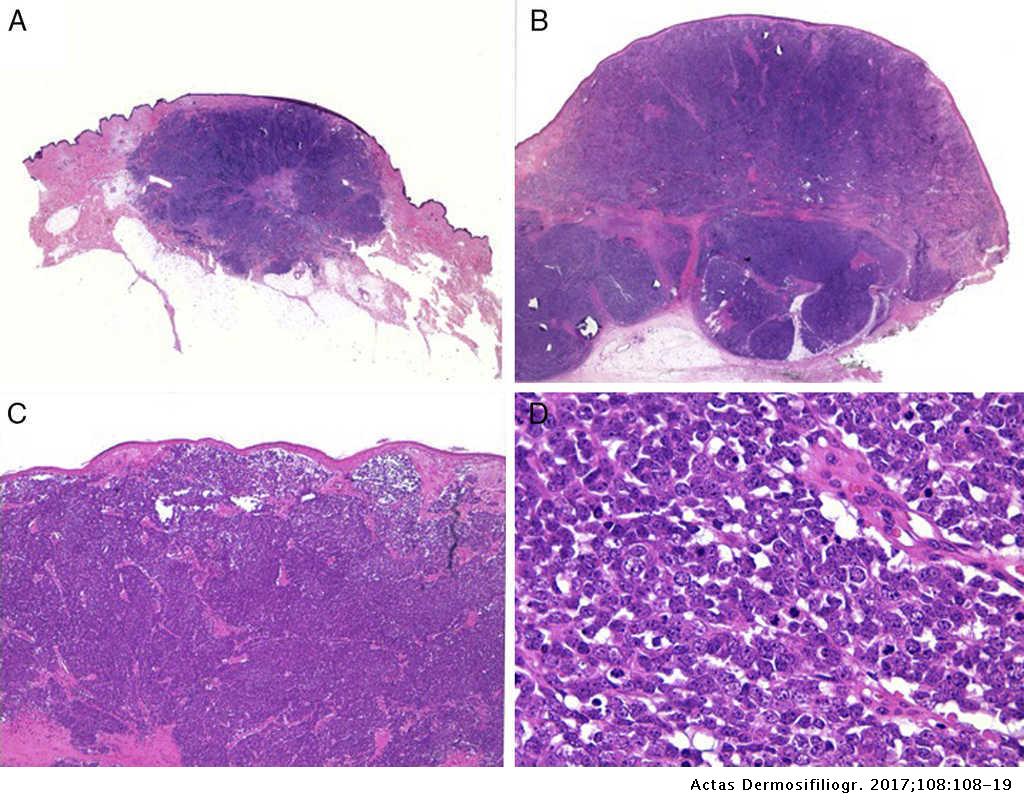
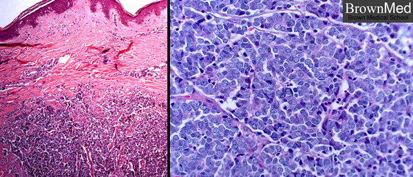
The tumour is centered in the dermis with frequent involvement of the overlying epidermis figures 1 2 and may invade the subcutaneous fat. Histology of merkel cell carcinoma. Merkel cells normally exist in the bottom basal layer of the epidermis about 01 mm from skins surface. The tumour forms sheets nests and rarely ribbons.
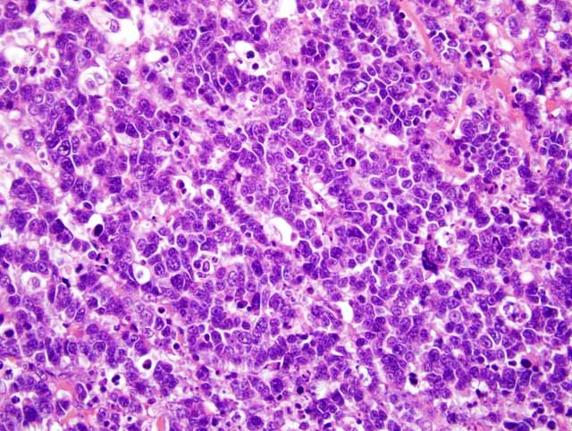
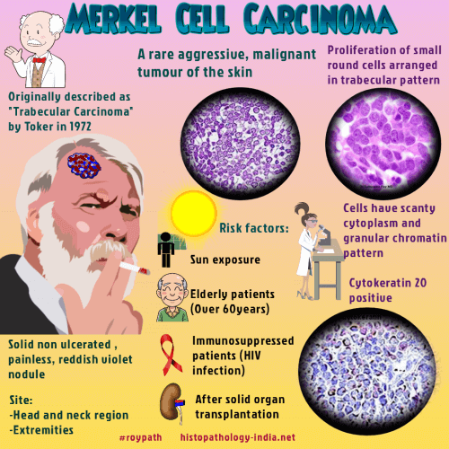
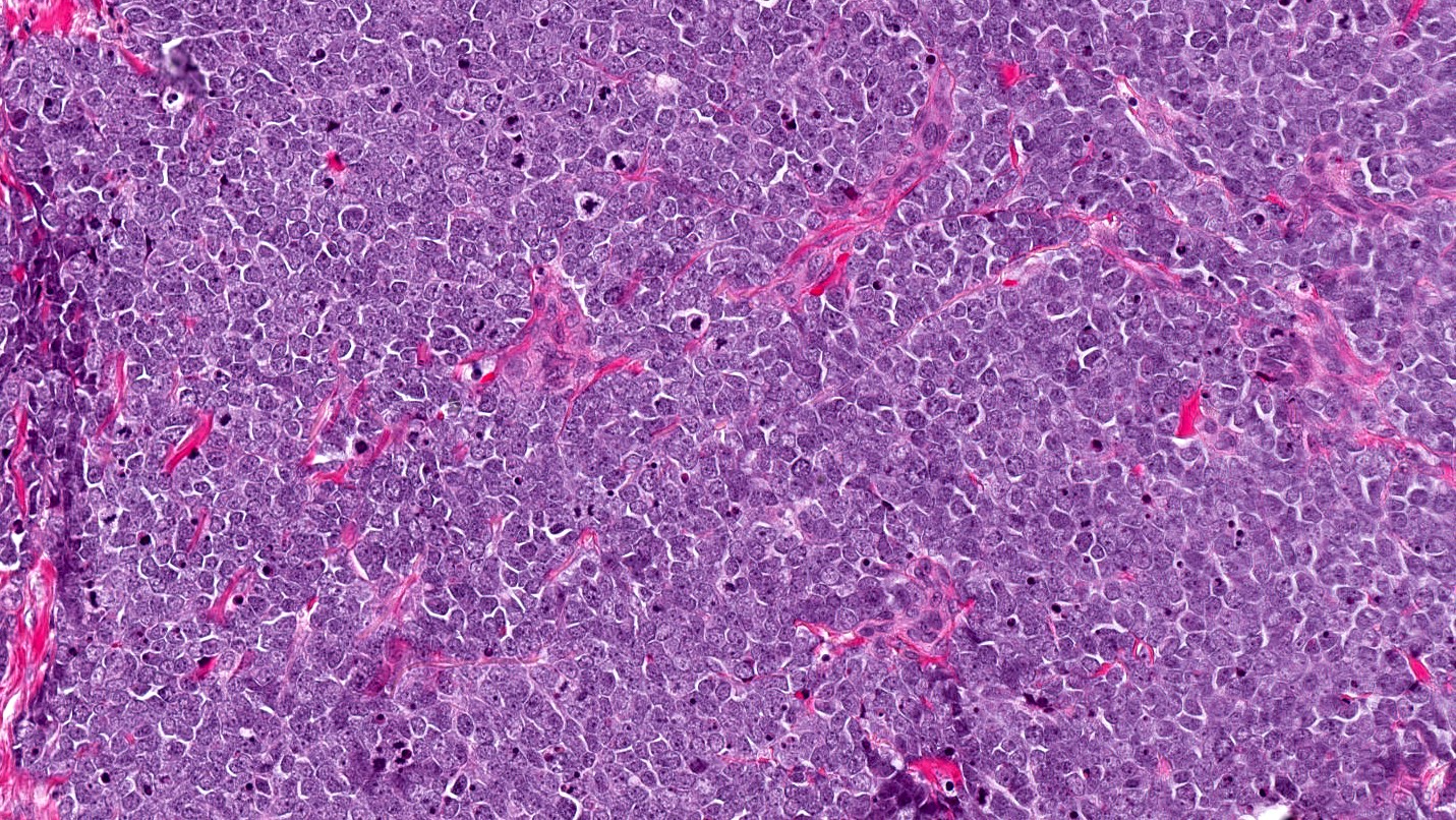
Merkel cell carcinoma is a neuroendocrine carcinoma composed of densely blue cells.
:background_color(FFFFFF):format(jpeg)/images/library/13197/skin-histology_med_mag_new_mag_boxes_english.jpg)



:background_color(FFFFFF):format(jpeg)/images/library/3470/7UckPYLm3F5EmutWIQmlQ_Meissner_Corpuscle.png)
