Epl Tendon Ultrasound
The tendon of epl is located over the back of the wrist where it bends through a tunnel adjacent to a slight prominence of bone.

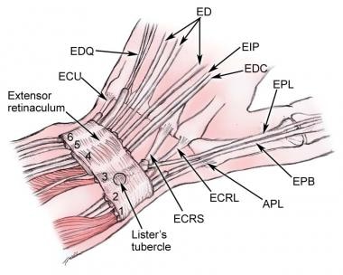
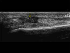
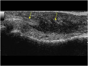
Epl tendon ultrasound. The extensor digitorum longus common tendon is adjacent in compartment 4. Extensor pollicis longus epl is a tendon which runs on the back side of the wrist and is confined to a tight tunnel. This tendon helps to straighten the end joint of the thumb and is essential to maintain hand span and to position the thumb in relation to the other fingers. Unfortunately this arrangement makes the tendon prone to damage from various causes and then it can rupture.
Investigations may include an x ray of the wrist and hand. The eip tendon is then re directed and sewn into the thumb bone or thumb tendon epl. This tendon is prone to damage and rupture due to its position and its utilization. Some patients may be able to feel a ping when the tendon ruptures.
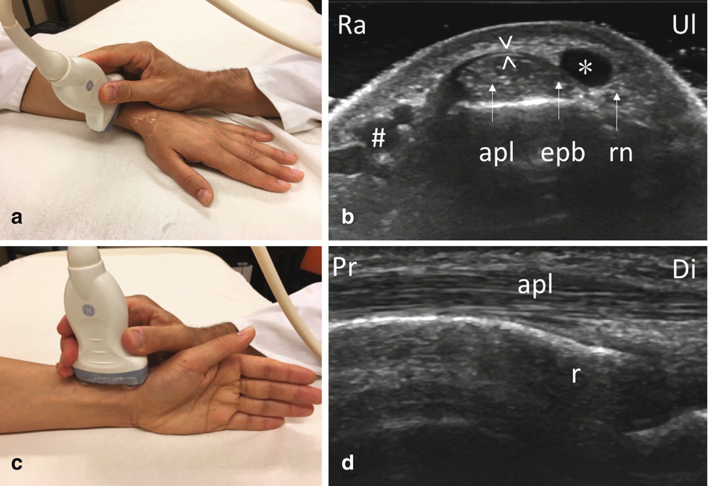
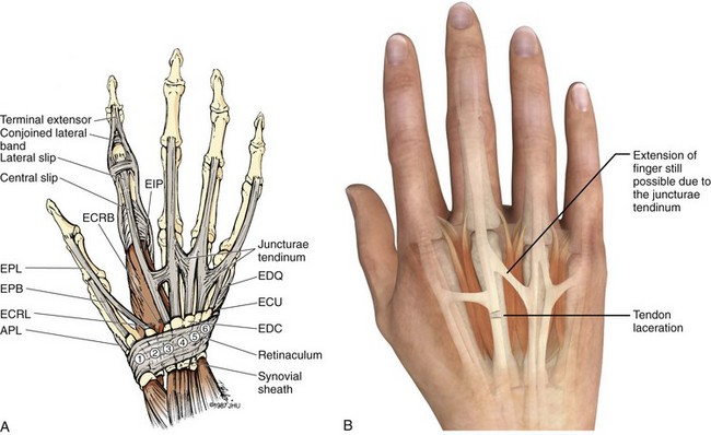
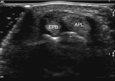
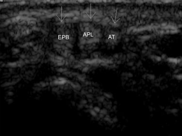
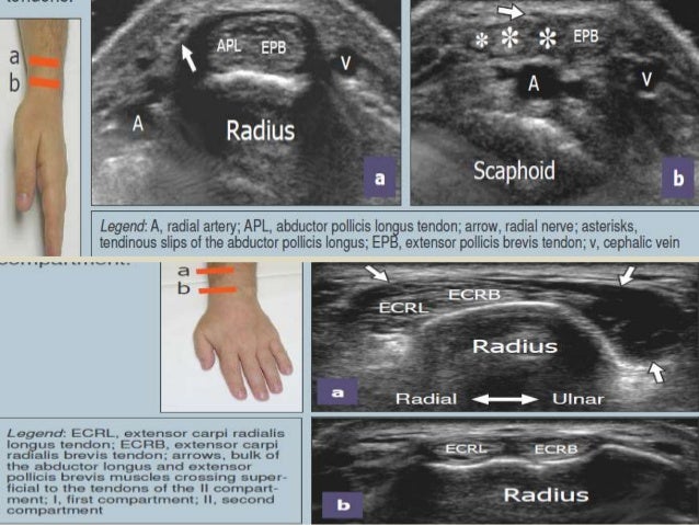
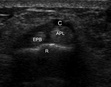
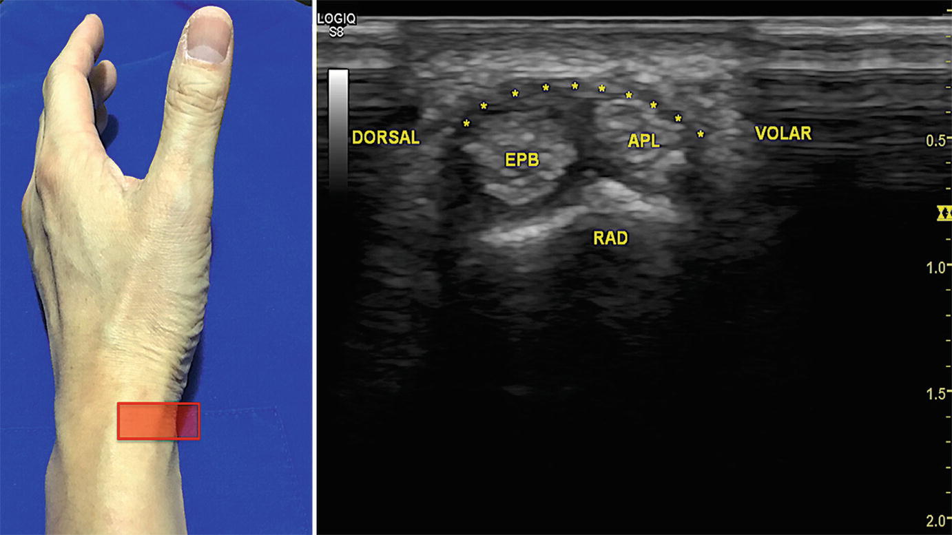
The common extensor digitorum tendon divides into 4 prior to the wrist crease. The abductor pollicis longus apl and extensor pollicis brevis epb ten. This oblique direction is so that it can pull the thumb up and back as if trying to hitchhike. On sonography the epl tendon was visualized in a transverse plane by rotating the probe 900 from the previously described position.
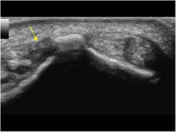
Common extensor digitorum with the overlying extensor retinaculum. Compartment 4 scan plane. Ultrasound is often used to confirm the rupture. The tendon insertion of the extra index finger extensor tendon extensor indicus proprius eip is detached.
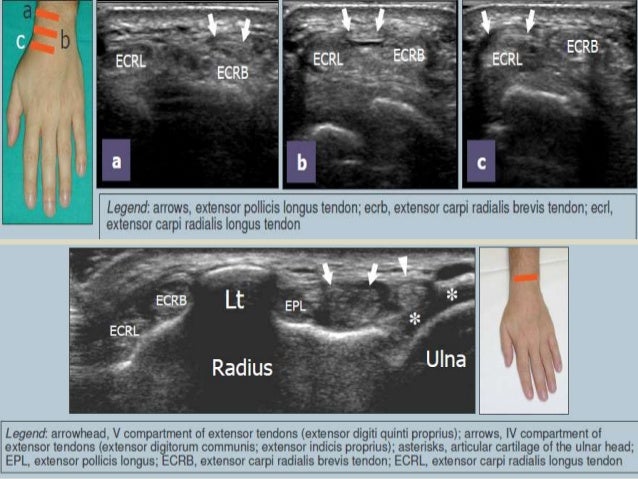
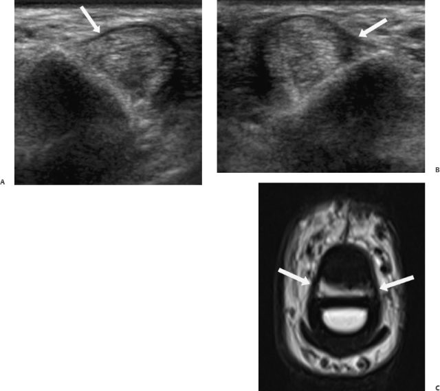
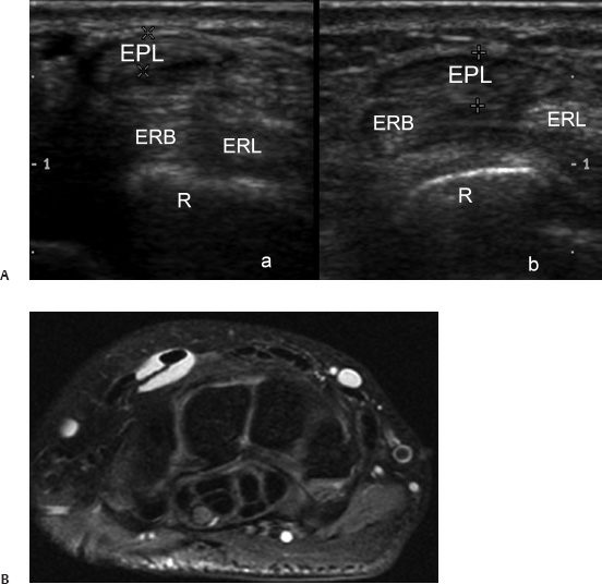
When the described position of the ultrasound probe is used along the transverse aspect of the tendon the epl tendon appears either hypoechoic or hyperechoic depending on the obliquity of the ultrasound beam. Transverse sonograms of the extensor surface of the wrist show the extensor digitorum ed and extensor pollicis longus epl tendons clearly and without artifact on the image obtained with the probe held exactly perpendicular to the tendons a but with a significant loss of echogenicity on the image obtained with the probe held at an oblique angle to the tendons b. Extensor pollicis longus rupture. Tendon runs through the third compartment figure 2c j ultrasound med 2016.
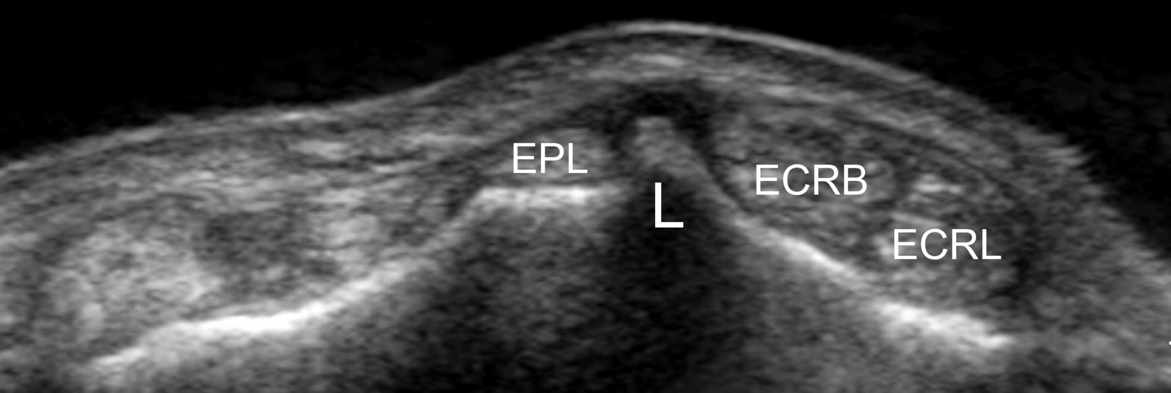
After this type of surgery a splint or cast is used for a few weeks after which supervised therapy may be started allowing gentle movement with a. The epl tendon is tucked against listers tubercle. What and where is the epl tendon. 3510811094 1083 gitto and draghinormal sonographic anatomy of the wrist figure 2.
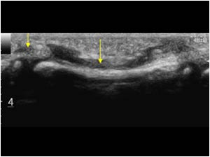
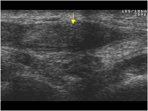
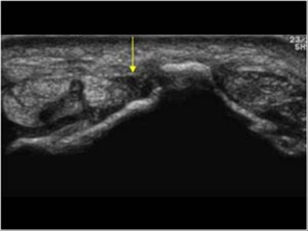
Dynamic ultrasound studies show normal movement of extensor pollicis longus epl tendon for passive flexion and ruptured epl on the right with 5 mm gap between the thickened ruptured ends.


