Epl Tendon Mri
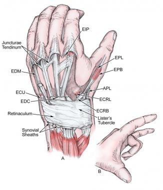
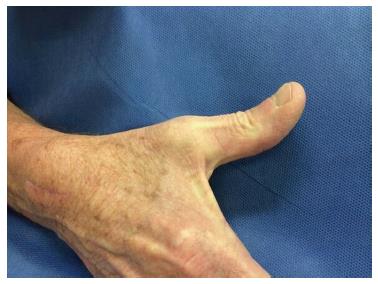
The tendon of epl defines the ulnar border of the anatomical snuff box.
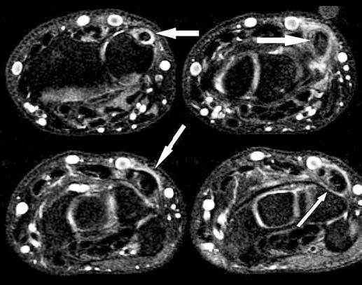
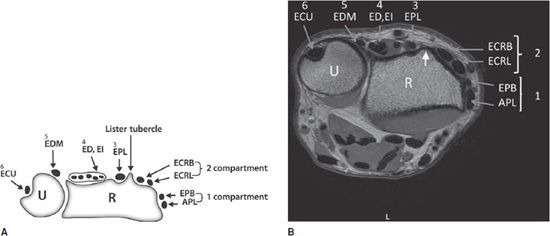
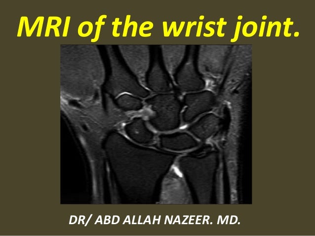
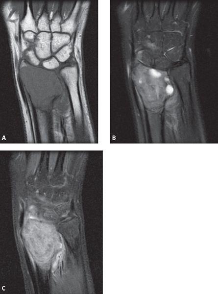
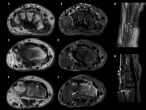
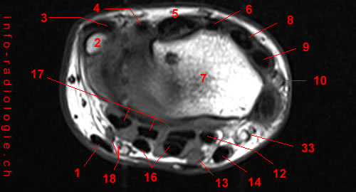
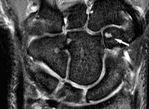
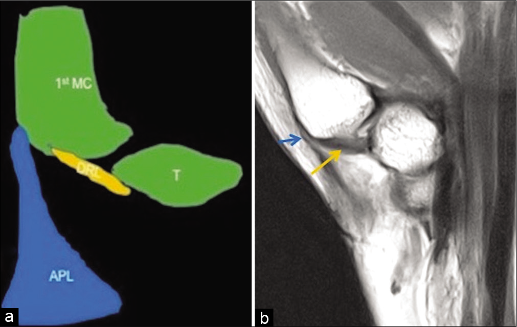
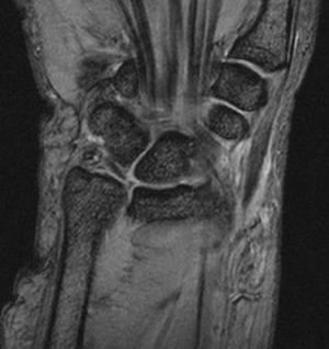
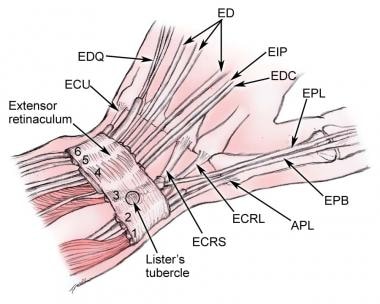
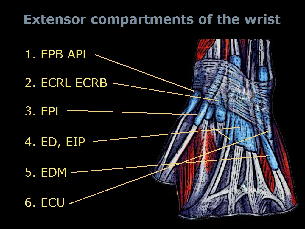
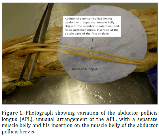
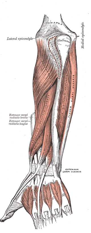
Epl tendon mri. This article reviews the normal anatomy of the extensor tendons of the wrist as well as the clinical presentation and mri appearances of common tendon abnormalities such as tears tenosynovitis intersection syndromes and associated or predisposing osseous findings. Mri findings are typical of tenosynovitis with fluid and high t2 signal intensity distending the tendon sheath of a thickened epl which frequently shows increased intrasubstance signal related to tendinosis 22 23. Extensor pollicis longus epl is a muscle of the deep compartment in the posterior compartment of the forearm. It is distinct from intersection syndrome which occurs more proximally in the forearm at the intersection of the first and second extensor compartments.
It passes through the 3 rd extensor compartment of the wrist then continues laterally towards the thumb around listers tubercle. The epl tendon is abnormally thickened with intermediate signal consistent with tendinosis. The distal intersection syndrome relates to tenosynovitis of the extensor pollicis longus epl tendon 3 rd extensor compartment where it crosses the extensor carpi radialis longus ecrl and brevis ecrb tendons 2 nd extensor compartment 1. Dr daniel j bell and greg mirt et al.


